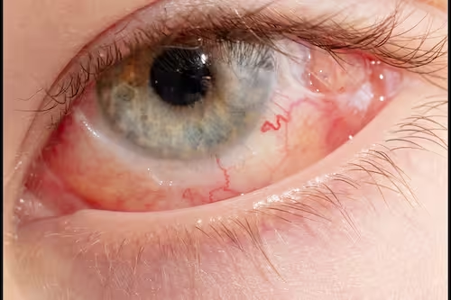
Detached Retina Treatment in DubaiSymptoms, surgery & recovery
A detached retina is a medical emergency: the light-sensitive layer at the back of the eye pulls away from the tissue that nourishes it.
It is usually painless. Without prompt surgery it can cause permanent vision loss, but repairs succeed in about 9 out of 10 cases.
Symptoms right now?
Sudden flashes of light, a shower of new floaters, or a dark shadow or curtain across your vision need same-day assessment. Call Ebsaar now; if the clinic is closed, go to the nearest hospital emergency department.
Medically reviewed by Prof. Dr. Hani Sakla, Consultant Ophthalmologist
·
Last reviewed
!3 warning signsFlashes, new floaters, a shadow or curtain.
9/10Repairs succeedin about 9 out of 10 cases (ASRS).
01FlashesSudden flashes of light, like a camera flash, often at the edge of vision.
02FloatersA sudden shower of new specks, threads or cobweb-like shapes.
03Shadow or curtainA dark area that spreads across part of your side vision.
04Blurred visionSudden blur or distortion, usually in one eye and without pain.
In 60 seconds
Detached retina, explained simply
What it is, how urgent it is and how it is repaired. The details and sources follow below.
1What it is
The retina lifts off
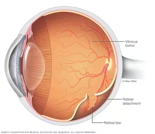
Fluid collects under the retina, usually through a tear, and separates it from the layer that supplies it with oxygen and nutrients.
Most common
Rhegmatogenous (caused by a tear)
Main causes
Age-related vitreous changes, high myopia, injury
Pain
Usually none
2How urgent
An emergency
The sooner the retina is reattached, especially before the central macula detaches, the better the chance of keeping good vision.
Act
Same-day examination
Tests
Dilated retinal exam, OCT, ultrasound
Heals alone?
No, it needs treatment
3How it is fixed
Retinal surgery
The retina is pushed back into place and the tear is sealed with laser or freezing treatment, usually under local anaesthetic.
Options
Pneumatic retinopexy, vitrectomy, scleral buckle
Success
About 9 in 10 repairs
Recovery
Blurry vision for 2–6 weeks
Direct answer
Can a detached retina be fixed?
Yes, in most cases. According to the American Society of Retina Specialists, retinal detachment repairs succeed in about 9 out of 10 cases, although some patients need more than one operation. How much vision returns depends mainly on how long the retina was detached and whether the macula, the centre of the retina, was involved. That is why speed matters.
01Don't waitSymptoms can come and go while the detachment spreads.
02Get a dilated retinal examSame day, by an ophthalmologist.
03Treat the tear or detachmentLaser for tears; surgery for detachments.
Key takeaways
It is an emergency. Sudden flashes, new floaters or a shadow in your vision need same-day assessment by an ophthalmologist.
It is usually painless. The lack of pain is one reason people delay, which can cost vision.
It does not heal on its own. Retinal tears are sealed with laser or freezing treatment; detachments need surgery.
Three main operations: pneumatic retinopexy, vitrectomy and scleral buckle, chosen by the size, location and type of detachment.
High success rate. Repairs succeed in about 9 out of 10 cases (ASRS); about 1 in 20 patients needs a second operation (AAO).
With a gas bubble in the eye, you must not fly or have nitrous oxide anaesthesia until your surgeon confirms the gas has gone.
What is a detached retina?
The retina is the thin, light-sensitive tissue that lines the back of the eye and converts light into signals the brain reads as vision. A detached retina (retinal detachment) happens when the retina separates from the layer beneath it, which supplies it with oxygen and nutrients. The detached part stops working, and without treatment the damage can become permanent.
A detachment can start in the side vision and spread towards the macula, the centre of the retina responsible for sharp reading and face vision. Retinal detachment affects roughly 1 in 10,000 people each year, and the risk rises with age, particularly after 50. Ebsaar’s retina service in Dubai examines suspected detachments urgently.
Types of retinal detachment
The U.S. National Eye Institute describes three types. Knowing the type helps the surgeon choose the right treatment.
The three types of retinal detachment
Type
What happens
Most common cause
Usual treatment
Rhegmatogenous
A tear or hole lets fluid pass under the retina. This is the most common type.
Age-related shrinking of the vitreous gel pulling on the retina.
Surgery: pneumatic retinopexy, vitrectomy or scleral buckle.
Tractional
Scar tissue on the retina’s surface pulls it away from the back of the eye.
Diabetic retinopathy.
Usually vitrectomy to remove the scar tissue.
Exudative
Fluid builds up behind the retina without any tear or break.
Leaking blood vessels or swelling, for example from inflammation or tumours.
Treating the underlying cause.
Swipe to compare →
Detached retina symptoms: the warning signs
Symptoms often start suddenly and can progress over hours or days. They usually affect one eye. If you notice any of the following, get an urgent eye examination even if your sight still seems fine:
Sudden flashes of light
Brief flashes, often at the edge of vision and more noticeable in dim light, can mean the vitreous is tugging on the retina.
New or many floaters
A sudden shower of dots, threads or cobweb-like shapes drifting across your vision.
Shadow or curtain
A dark or grey shadow spreading across part of your side vision is the classic sign that the retina has detached.
Blurred or distorted vision
Straight lines may look wavy, or vision may become hazy if the detachment reaches the macula.
No pain
A detached retina usually does not hurt. Pain is not a reliable warning sign, so never wait for it.
Early examination gives the best chance of keeping the macula attached and protecting central vision.
When is it an emergency?
Treat these as an eye emergency and seek same-day care from an ophthalmologist or a hospital emergency department:
Flashes or floaters that appear suddenly or increase suddenly.
A shadow, curtain or dark area moving across your vision.
Sudden blurred or distorted vision in one eye.
Any of these after an eye injury, or after cataract or other eye surgery.
Symptoms in one eye if you have had a retinal tear or detachment before.
Causes and risk factors
Most detachments start with a retinal tear. As we age, the vitreous gel that fills the eye shrinks and can pull away from the retina, sometimes hard enough to tear it. Fluid then passes through the tear and lifts the retina. You are at higher risk if you:
Are highly nearsighted (high myopia), because a longer eye has a thinner, more stretched retina.
Have had cataract surgery or other eye surgery.
Have had an eye injury, for example from contact sports or an accident.
Have had a retinal tear or detachment before, in either eye.
Have a family history of retinal detachment.
Have lattice degeneration (thin, weak areas of the retina) or diabetic retinopathy.
Retinal tear vs retinal detachment
A retinal tear is a break in the retina that has not yet let enough fluid underneath to lift it. Found early, a tear can often be sealed in the clinic with laser photocoagulation or cryotherapy (freezing), which create a scar around the tear and can prevent a detachment. A retinal detachment means the retina has already lifted, and it needs surgery to reattach it. This is why new flashes and floaters should be checked quickly, before a tear becomes a detachment.
Diagnosis
How a detached retina is diagnosed
Diagnosis is made by an ophthalmologist during a same-day examination. It is quick and painless.
01
Dilated retinal exam
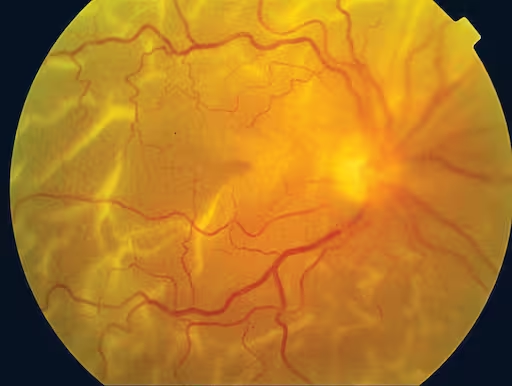
Drops widen the pupil so the doctor can examine the whole retina with special lenses and find every tear.
02
OCT scan
Optical coherence tomography gives cross-section images showing whether fluid has reached the macula.
03
Ultrasound
Used when bleeding or a cataract blocks the view, to see whether the retina is attached.
Type: tear-related, tractional or exudative.
Macula: still attached (macula-on) or detached (macula-off).
Tears: number, size and position of breaks.
Extent: how much of the retina has lifted.
Scar tissue: any proliferative changes that affect the repair.
Other eye: checked for tears or weak areas too.
Detached retina treatment options
Treatment depends on the type, size and location of the detachment, the number of tears and whether scar tissue is present. Surgery is usually done under local anaesthetic, so you are awake but the eye is numbed, and most people do not need to stay in hospital overnight (NHS).
Detached retina surgery compared
Feature
Pneumatic retinopexy
Vitrectomy
Scleral buckle
How it works
A gas bubble injected into the eye presses the retina back; the tear is sealed with laser or freezing.
The vitreous gel is removed and replaced with gas, air or silicone oil to hold the retina in place.
A soft silicone band sewn to the outside of the eye indents the wall towards the retina.
Often used for
Small, uncomplicated detachments with a tear in the upper retina.
Larger or complex detachments, including those with scar tissue or bleeding.
Selected detachments, often in younger patients; can be combined with vitrectomy.
Incision
Minimal: an injection only
Small ports in the white of the eye
Band placed on the outside of the eye
Head positioning
Yes, for several days, when a gas bubble is used.
Usually not needed unless combined with gas.
Main trade-off
Strict positioning is essential for success.
Speeds up cataract formation; silicone oil needs a second operation to remove.
The band usually stays in place and can change the eye’s prescription.
Swipe to compare →
Pneumatic retinopexy
A small gas bubble is injected into the eye. You then hold your head in a set position so the bubble floats against the tear and presses the retina back into place, and the tear is sealed with laser or cryotherapy. It avoids a major incision and can be done in the clinic, but strict positioning is critical.
Vitrectomy (pars plana vitrectomy)
The surgeon removes the vitreous gel that is pulling on the retina, flattens the retina, seals the tears with laser and fills the eye with gas or silicone oil. A gas bubble is absorbed over several weeks, depending on the gas used; silicone oil is usually removed in a later operation. Vitrectomy is used for more complex detachments, including those with scar tissue.
Scleral buckle
A flexible silicone band is stitched to the sclera, the white outer wall of the eye, gently pushing the wall inwards so it meets the detached retina. The band usually stays in place permanently. It has been used for decades and can be combined with vitrectomy.
Your surgeon explains which operation suits your detachment and what recovery involves.
Recovery after detached retina surgery
Recovery depends on the operation, but most people eventually return to their normal activities. This is a typical course based on NHS guidance; your surgeon gives you a personal aftercare plan.
First daysThe eye may be sore and red; eye drops prevent infection and inflammation. If you have a gas bubble, you keep the head position your surgeon prescribes.
2–6 weeksVision is often blurry, and you may be advised not to drive. Do not fly while there is gas in the eye. Avoid heavy lifting and strenuous exercise until your surgeon agrees.
Several monthsVision continues to improve as the retina recovers. Your glasses prescription may change and is usually updated once the eye has settled.
Gas bubble safety rules
No flying or high-altitude travel until your surgeon confirms the gas has fully absorbed. The bubble expands at altitude and can raise eye pressure dangerously.
No nitrous oxide (“laughing gas”) anaesthesia. Tell every doctor and dentist you have gas in your eye, and wear the wristband your surgeon gives you.
No scuba diving or mountain trips until you are cleared.
Keep the prescribed head position. It keeps the bubble pressing on the tear while it heals.
Risks of detached retina surgery
All surgery carries risks, but untreated retinal detachment usually leads to permanent vision loss in that eye. Your surgeon will discuss:
Re-detachment: about 1 in 20 patients needs a second operation (AAO).
Cataract formation, which vitrectomy can speed up.
Raised eye pressure that needs monitoring or treatment.
Bleeding inside the eye, which usually settles.
Infection, which is rare.
Scar tissue (proliferative vitreoretinopathy) that can pull the retina off again.
Detached retina surgery cost in Dubai
The cost depends on the operation needed (pneumatic retinopexy, vitrectomy or scleral buckle), whether gas or silicone oil is used, any second procedure such as oil removal, and the follow-up included. Ebsaar explains the treatment plan and cost after the examination. Retinal detachment repair is a medically necessary operation, so ask your insurer about cover and pre-approval, but never delay urgent treatment while waiting for paperwork.
Ebsaar · Dubai
Retina care at Ebsaar Eye Surgery Center
Ebsaar Eye Surgery Center in Jumeirah, Dubai provides comprehensive ophthalmology care, including retina services, cataract surgery, glaucoma management, cornea treatment and vision correction. If you have symptoms of a retinal tear or detachment, call the clinic so your examination can be arranged without delay.
Urgent assessmentDilated retinal examination and imaging to confirm the diagnosis.
Treatment planningLaser for tears, and surgery options explained for detachments.
Jumeirah, DubaiVilla 32A, Jumeirah Beach Road, Dubai, United Arab Emirates.
Flashes, floaters or a shadow?
Don’t wait for it to hurt. Get your retina checked today.
Call Ebsaar Eye Surgery Center in Dubai to arrange an urgent retina examination. If the clinic is closed, go to the nearest hospital emergency department.
Short, direct answers to the questions patients ask most. They do not replace an eye examination.
What does vision look like with a detached retina?
Most people notice sudden flashes of light, a shower of new floaters, or a dark shadow or curtain spreading across part of their side vision. Vision can also become blurred or distorted, usually in one eye. The more of the retina that detaches, especially the central macula, the more vision is affected.
What does a detached retina feel like?
A detached retina usually does not hurt. The signs are visual, not physical: flashes, floaters, a shadow or blurred vision. Because there is no pain, people sometimes delay getting help, which increases the risk of permanent vision loss.
What causes a detached retina?
Most detachments start with a retinal tear. With age, the vitreous gel inside the eye shrinks and can pull on the retina hard enough to tear it, letting fluid pass underneath. High myopia, previous eye surgery such as cataract surgery, eye injury, a previous retinal tear and a family history all increase the risk.
Can a detached retina heal on its own?
No. A detached retina does not reattach by itself and needs treatment. A retinal tear found before it detaches can often be sealed with laser or freezing treatment, but a detachment needs surgery.
Can a detached retina be fixed?
Yes, in most cases. Retinal detachment repairs succeed in about 9 out of 10 cases, according to the American Society of Retina Specialists, although some people need more than one operation. How much vision returns depends on how long the retina was detached and whether the macula was involved.
How quickly does a detached retina need treatment?
As soon as possible. Sudden flashes, floaters or a shadow need a same-day eye examination. Detachments that have not yet reached the macula are treated urgently to protect central vision, because the longer the retina stays detached, the higher the risk of permanent loss.
Can a detached retina cause blindness?
Yes. Without treatment, a detached retina usually leads to permanent vision loss in the affected eye. Prompt surgery gives the best chance of saving sight, although some vision may remain reduced if the macula was detached.
Is detached retina surgery painful?
Surgery is usually done under local anaesthetic, so you are awake but the eye is numbed and you should not feel pain. Afterwards the eye may be sore and red for a few days, which is managed with drops and simple pain relief.
How long is recovery after retinal detachment surgery?
Vision is often blurry for 2 to 6 weeks, and the eye may be sore and red. You may not be able to drive during this time. Vision can keep improving for several months, and most people eventually return to their normal activities.
Can I fly after retinal detachment surgery?
Not while there is a gas bubble in your eye. The gas expands at altitude and can dangerously raise eye pressure. Your surgeon will tell you when the gas has fully absorbed and it is safe to fly. Silicone oil does not carry the same flying restriction.
How common is a detached retina?
Retinal detachment is relatively uncommon, affecting roughly 1 in 10,000 people each year. The risk rises with age, especially after 50, and is higher in people who are very nearsighted, have had eye surgery or an eye injury, or have a family history.
Can the symptoms of a detached retina come and go?
Yes. Flashes and floaters can seem to come and go, but that does not mean the problem has resolved. A tear or small detachment can spread over hours or days, so any new symptoms should be checked urgently.
Can a detached retina cause double vision or dizziness?
Not directly. A detached retina does not usually cause true double vision or dizziness, but blurred or distorted vision in one eye can feel disorienting. Any sudden change in vision should be examined promptly.
How much does detached retina surgery cost in Dubai?
The cost depends on the operation needed, whether gas or silicone oil is used, any further procedures and the follow-up included. Ebsaar explains the treatment plan and cost after the examination. Ask your insurer about cover, but do not delay urgent treatment.